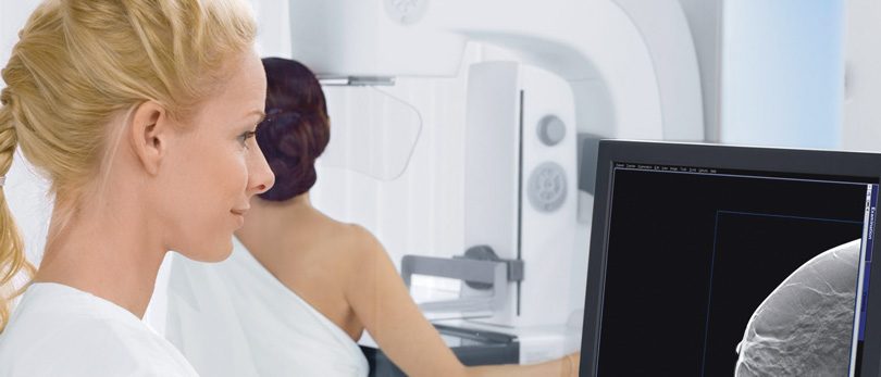
The newest technology in mammography is making great strides toward eliminating the need for unnecessary diagnostic mammograms and may find more invasive breast cancers. This new technology is now available at the UHS Breast Center. The technology is called digital breast tomosynthesis, more commonly known as 3-D mammography.
Mammograms remain the first line of defense in the early detection of breast cancer. However, regular mammograms do have limitations. Regular screening mammograms basically show a flat, 2-dimensional image of the entire compressed breast.
With some women — especially those with very dense breasts — it’s often difficult for radiologists to determine if certain anomalies on mammograms are malignancies, or just normal breast tissue that is too dense to see clearly. This leads to a significant number of “false positives,” meaning there is enough evidence of a malignancy to require a patient to undergo additional diagnostic imaging, but the anomaly is eventually determined to be benign.
However, now that mammograms use digital imaging (instead of film), dozens of images of the breast can be taken from several different angles, and then synthesized through the computer using tomosynthesis to create a three-dimensional image of the breast. This allows doctors to see inside breast anomalies with far more clarity.
“The biggest advantage of tomosynthesis is that it reduces or eliminates the need for additional diagnostic mammograms, which is often a point of anxiety for patients and their families,” says Michael J. Farrell, MD, breast surgeon at UHS. “When a patient gets a call that her mammogram was ‘suspicious’ and she needs to come back to the center for additional imaging procedures, it’s very stressful. Tomosynthesis allows us to compile a host of one-millimeter slices of breast images from different angles at once. This level of clarity often reduces or eliminates the need for additional imaging.”
“However, while 3-D mammograms can save time and stress — not to mention unnecessary radiation — studies on the overall effectiveness of 3-D mammograms in finding breast cancer are conflicting,” says Dr. Farrell. “Several studies have affirmed the superiority of 3-D over 2-D imaging, but some have not shown this.”
Still, reducing false positives, expense and the stress that goes along with additional unnecessary testing is reason enough to be excited about this new technology.
Currently, the 3-D technology at the UHS Breast Center is used primarily for diagnostic mammograms. However, as more machines and capacity are added over the next year, additional groups of patients who may benefit from this technology will be advised to take advantage of it. These decisions are driven by clinical indications and collaboration between referring providers and the UHS Breast Center team.
GET A CLEAR PICTURE.
If you have questions, talk to your doctor or call the UHS Breast Center at 763-5523.


